Article:
Emphasizing the importance of words, we pronounce them slowly, and something secondary and insignificant - as if in a “patter.” However, there are speech pathologies in which the pronunciation of words is so accelerated or slow that the understanding of speech is lost - it is so illegible. These are signs of speech pathology - disturbances in the tempo and rhythm of speech. An adult and a child with such a pathology, seeing how others react to his speech, tries to speak out less so as not to get into an awkward position. What types of speech tempo and rhythm disorders are there? How to diagnose such disorders? How to work with such children in a regular school?
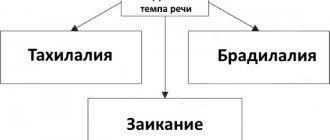
Types of speech rhythm and tempo disorders
Not only a speech therapist, but also parents and teachers can hear that a child’s speech deviates from the normal pace and the rhythm is disturbed, but only a specialist can determine the structure and cause of the defect.
Violations of the tempo and rhythm of speech include:
- Tahilalia is the accelerated pronunciation of words, sounds, syllables at a speed of 20-30 sounds per second (instead of 9-14 normally). The rapidity of speech negatively affects the articulation of sounds. Pathology occurs in childhood, becomes established during adolescence, and can remain with a person for life.
- Bradylalia is a pathologically slow speech, most often found in children with mental retardation or mental retardation, since violations of basic mental processes (memory, attention, thinking) and fine motor skills are often observed in parallel with bradylalia.
- Stumbling - against the background of accelerated speech, repetition of words, syllables and sounds occurs. It must be distinguished from stuttering, since stumbling does not have convulsions of the speech apparatus in its etiology; a child or adult with stumbling does not experience fear of speaking.
- Stuttering - during a statement, involuntary stops occur that are beyond the control of the stutterer. They are accompanied by convulsions of all parts of the speech apparatus.
Tips for parents
It is important for the child to create a positive speech environment.
Repeatedly talk through ordinary, daily activities:
dressing, washing, eating, bathing, walking, getting ready for bed, going to bed, cleaning the room and toys, cooking. All these processes must necessarily be accompanied by the speech of adults.
Say nursery rhymes, tongue twisters, proverbs, proverbs, sing songs
.
Talk to your child as often as possible
, listen carefully to his answers to questions. This will not only improve your baby’s speech, but will also establish a trusting relationship between you.
Read together
, discuss what you read, illustrate the plot together, discuss the drawings.
Symptoms of speech tempo and rhythm disorders
With tachylalia (accelerated speech rate), the rapid flow of utterance stuns the interlocutor, whom the speaker practically does not listen to the end of, and often interrupts. The articulatory apparatus does not have time to clearly pronounce sounds, syllables and words; the entire utterance is pronounced in one exhalation, without respite. Tachylalia may be accompanied by twitching of the body or separately of the arms and legs, and grimacing.
Stumbling is distinguished, in addition to the frequent repetition of speech units, by unjustified pauses in speech, stops in the middle of a speech utterance. Unlike stuttering when stumbling, a child’s speech does not deteriorate in front of an unfamiliar audience and does not have periodicity.
With bradylalia , the articulation of sounds is sluggish and unclear, vowel sounds are pronounced stretched out. The voice is monotonous, the statements are not emotionally colored, they lack characteristic modulations. Speech is so slow that it causes impatience and irritation among others.
Children, seeing such an attitude, try to remain silent more often than to speak. This position further retards their mental development. According to scientific research, with bradylalia, not only external, but also internal speech has a slow pace.
Stuttering has a wide clinical picture. It is characterized by the following symptoms:
- Convulsions of all parts of the articulatory apparatus.
- Accompanying movements of the muscles of the face, neck, arms and legs - squinting, stomping, clenching fists, frequent blinking.
- Speech tricks - all kinds of “uh”, “well”. “so”, “here”, etc.
- Fear of speech, fear of pronouncing individual sounds and words.
- Increased stuttering when communicating with unfamiliar people.
Classification of cortical dysarthria in children
Depending on the location of the pathological focus and the mechanism of development, two types of pathology are distinguished:
- Cortical kinetic dysarthria (efferent) is associated with damage to the central zone of the anterior cortex. Due to the proximity of the area that controls hand movements, hemiparesis may occur. Characterized by slowness of speech and lack of rhythm. Some children speak in syllables;
- Kinesthetic (afferent) dysarthria is associated with damage to the postcentral zones. It is characterized by discoordinated work of the organs of articulation and movements of the muscles of the hand on the opposite side.
Based on severity, there are 4 forms of the defect. In the first degree there is no clear clinical picture, then their severity increases. Grade 4 may be accompanied by complete anarthria, that is, lack of speech.
Mixed variants of the disease are possible, for example, cortical-subcortical dysarthria, when in addition to the cortical part, the subcortical parts of the brain are also affected.
Examination of children with violations of the tempo and rhythm of speech
If parents suspect that their child has disturbances in the tempo and rhythm of speech, they should consult with a speech therapist at a children's clinic, speech center, school or kindergarten. Sometimes you can hear the erroneous opinion that you should not start correcting speech deficiencies until the child is 5 years old.
This is partly true for uncomplicated cases of sound pronunciation formation, since the speech apparatus has not yet been formed, and sounds can appear on their own. In all other cases, the sooner correction work begins, the more successful it will be.
At the initial visit, the specialist is required to conduct a speech therapy examination. The main directions of this survey:
- Establishing the cause and nature of speech impairment.
- Determining the rate of speech of the child and his parents.
- Diagnosis of basic mental processes (memory, attention, thinking).
- Studying the history of speech development - the appearance of the first words, phrases, the likelihood of parents forcing the child’s speech. Bilingualism in the family.
- Determining the style of family education, attitude towards the child, the presence of conflicts, difficult life situations.
- Collecting anamnesis of the disorder and concomitant pathologies.
- Determination of existing articulation deficiencies.
- Correspondence of the level of speech development of the child to the age norm.
Perhaps some questions may seem unnecessary to parents, but the speech therapist does not ask them out of idle curiosity - there are no trifles in the development and correction of speech disorders, the situation in the family and the presence of psychological problems are very important.
Speech Disorder Specialists
Speech therapist.
Most speech disorders are a speech therapy problem, which is why speech therapists should be contacted first.
Neurologist
provides medication support to children with cerebral palsy, alalia, dysarthria, etc.
A defectologist is able to significantly “pump up” a child’s brain, develop intelligence and thinking.
An otorhinolaryngologist determines pathologies of the peripheral speech apparatus of a medical nature: hearing loss, cleft palate, adenoid growths, sore throat, etc.
A teacher of the deaf is involved in the development of speech in deaf and hard of hearing children and adults.
Psychologist. Due to shyness or aggressive behavior, the child’s speech is distorted and impoverished. Problems in the family and with peers can cause delayed speech development.
Neuropsychologist (clinical psychologist) - in case of speech disorders (caused by impaired brain function), teaches the child’s brain to function correctly through motor and play exercises.
| Defectology and speech therapy, as well as Neurology services are available in our center. |
Causes of speech pathologies
The basis for disturbances in the tempo and rhythm of speech is an incorrect relationship between the processes of excitation and inhibition in the cerebral cortex.
The causes of tachylalia (accelerated speech rate) are:
- The predominance of excitation processes in the cerebral cortex, responsible for the formation of speech.
- Heredity, congenital characteristics of temperament.
- Imitating the fast-talking of others, mistakes in education - such speech is most often found in unbalanced and excitable children.
The causes of bradyllalia (slow speech) can be:
- Dominance of inhibition over excitation in the speech areas of the cerebral cortex.
- This feature of external and internal speech can be inherited.
- Pathology appears as a result of a child’s imitation of incorrect speech patterns from others.
Causes of stuttering can be:
- Unbearable speech load (learning large poems, memorizing texts that are not age appropriate, memorizing difficult and obscure words).
- Frequent punishment of children, sudden and severe fear, mental trauma due to improper upbringing.
- Excessively accelerated speech due to imitation of family members, getting stuck on individual sounds during speech;
- Traumatic brain injuries, neuroinfections.
The main factors provoking pathology of speech development are congenital or acquired weakness of the nervous system and a decrease in its stability.
ARRHYTHMIAS: diagnosis and treatment
Arrhythmia (heart rhythm disturbance) is one of the most common pathologies (15-25%) in the practice of a cardiologist. Arrhythmia threatens not only the health, but, in some cases, the life of the patient. It is known that 10% of patients who survive acute myocardial infarction (MI) die within a year. The main risk factors for sudden death after MI are ventricular arrhythmias and heart failure (HF).
Cardiac arrhythmias are a change in the basic electrophysiological properties of the heart (automatism, excitability, conductivity), leading to a violation of the coordinated contraction of the entire heart or its parts and manifested by a change in the frequency and regularity of the heart rhythm.
It is very important for the doctor to establish the underlying cause of the disease that caused the arrhythmia. Sometimes this is associated with certain difficulties, since the causes of arrhythmia can be different: extracardiac, cardiac and idiopathic (primary electrical heart disease).
Extracardiac factors for the development of arrhythmia include functional and organic lesions of the central nervous system, dysfunction of the autonomic nervous system, endocrine diseases, electrolyte imbalance, mechanical and electrical trauma, hypo- and hyperthermia, excessive physical activity, intoxication with alcohol, nicotine, coffee, and drugs. This is especially true for sympathomimetics, cardiac glycosides, diuretics, many psychotropic and other drugs, including antiarrhythmics.
Cardiac factors are primarily coronary heart disease, heart defects, heart failure, arterial hypertension, inflammatory and non-inflammatory myocardial lesions, diagnostic manipulations and operations on the heart and coronary vessels.
Today there is a European standard for the diagnosis and treatment of arrhythmias. If arrhythmia is suspected, the doctor must establish the fact of rhythm disturbances, determine the nature of the arrhythmia, its cause, functional or pathological nature, and decide on the use of antiarrhythmic therapy. For these purposes, physical examinations, ECG, daily ECG monitoring (Holter), esophageal electrocardiography are used.
To clarify the diagnosis use:
- long-term recording of leads II, aVF, doubled ECG voltage and increased paper tape speed to 50 mm/s to identify P waves;
- additional ECG leads, ECG recording during carotid sinus massage for 5 s, bolus tests with medications and exercise tests.
According to the clinical classification, arrhythmias are divided according to the functional principle: dysfunction of automaticity, excitability, conduction and combined arrhythmias. Automatic dysfunctions include sinus tachycardia, bradycardia, arrhythmia and migration of the rhythm source; dysfunction of excitability - extrasystole, paroxysmal and non-paroxysmal tachycardia, flutter and fibrillation (fibrillation) of the atria and ventricles. Conduction disorders include blockades: sinoauricular, atrial, atrioventricular, intraventricular; ventricular asystole. And the most complex are combined arrhythmias: sick sinus syndrome, escape contractions and rhythms, atrioventricular dissociation, premature ventricular excitation syndrome and parasystole.
All tachyarrhythmias are divided into two types.
- Tachycardia with a narrow QRS complex (antegrade conduction through the AV node); most often it is supraventricular paroxysmal tachycardia. It is treated carefully by administering verapamil, propranolol or digoxin intravenously.
- Wide QRS tachycardia (antegrade conduction through the accessory pathway) is often associated with atrial fibrillation and a very high (>250 beats/min) ventricular rate. If hemodynamic parameters are unstable, immediate cardioversion is indicated; Drug treatment is carried out with lidocaine or procainamide intravenously.
Arrhythmias with a narrow QRS complex
- Atrial extrasystole is an altered wave with normal QRS width. If symptomatic, β-blockers or group IA drugs are prescribed.
- Sinus tachycardia - heart rate 100–160 with a normal P wave. First of all, the causes of arrhythmia are eliminated, if the arrhythmia is symptomatic - β-blockers.
- Paroxysmal supraventricular tachycardia - at a heart rate of 140–250, the P wave is pointed or inverted in leads II, III, aVF. Adenosine, verapamil, β-blocker, group IA drug, and electrical pulse therapy (150 J) are prescribed. Paroxysmal atrial tachycardia with block - at a heart rate of 130–250, a straight pointed P 2:1, 3:1, 4:1 block. If digitoxin was administered, it should be interrupted and K+ corrected; diphenine 250 mg IV over 5 minutes.
- Atrial flutter - heart rate 250–350, saw-tooth flutter waves, 2:1, 4:1 T blockade. Digoxin, β-blockers or verapamil are administered to reduce the ventricular rate.
- Atrial fibrillation - heart rate>350, P is indistinguishable, QRS intervals are irregular. EIT may be required to restore sinus rhythm (after prolonged anticoagulation) with procainamide or quinidine (flutter: 50 J; fibrillation: 100-200 J).
- Multifocal atrial tachycardia - with a heart rate of 100–220 - more than three differentiated forms of the P wave with different P-P intervals. If there is pathology of the lungs, treatment of the underlying disease is required, verapamil is prescribed to reduce the frequency of ventricular contractions.
Arrhythmias with wide QRS complex
- Ventricular extrasystoles - with complete compensatory pauses between normal complexes. The use of certain drugs, as for ventricular tachycardia.
- Ventricular tachycardia is a moderately severe irregularity with a heart rate of 100–250. In case of unstable hemodynamics, cardioversion (100 J) or intravenous administration of procainamide, lidocaine, or bretylium is indicated. For long-term prevention, it is advisable to prescribe drugs of groups IA, IB, 1C, III.
- Ventricular tachycardia of the “pirouette” type is sinusoidal oscillations with a height in the QRS. The administration of lidocaine is indicated, in the absence of a history of coronary heart disease - isoproterenol, bretylium, magnesium. Quinidine and all drugs that prolong the QT interval are contraindicated.
- Ventricular fibrillation - unstable electrical activity on the ECG. Immediate defibrillation is needed (200-400 J).
- Supraventricular tachycardia with aberrant ventricular conduction - a wide QRS complex with a P wave typical of a supraventricular rhythm.
Treatment is prescribed as for the corresponding supraventricular rhythm, but if the ventricular rate exceeds 200 - as for WPW syndrome.
Atrioventricular block
I degree. An extended constant PR interval (0.20 s) does not require treatment and can be considered as normal or be caused by vagotonia, which often occurs after the use of digitalis.
II degree. Mobitz I (Wenckebach). The ECG shows a narrow QRS complex with a progressive increase in the PR interval until the QRS complex drops out, after which the sequence is repeated. It is observed with digitalis intoxication, vagotonia, and lower myocardial infarction. Usually does not require treatment; if the arrhythmia is symptomatic, atropine 0.6 mg is injected into a vein (the injection is repeated three to four times). In some situations, a temporary pacemaker is used.
Mobitz II. The PR interval is fixed, the complexes periodically fall out in a ratio of 2:1, 3:1 or 4:1, the QRS complex is wide. Usually occurs with damage to the conduction system or myocardial infarction. This rhythm is life-threatening and often leads to complete AV block. To eliminate the pathology, a pacemaker is prescribed.
III degree (complete AV block). The atria and ventricles contract independently of each other. It is observed in cases of damage to the conduction system of the heart, myocardial infarction, and digitalis intoxication. If there is no transient combination of complete AV block with asymptomatic congenital heart block or inferior myocardial infarction, a permanent pacemaker is indicated.
Pre-excitation syndrome (WPW)
The impulse is carried out through an additional pathway between the atria and ventricles. The ECG is characterized by a short PR interval and merged, upward-directed QRS complexes (delta wave).
At the treatment stage, it is important to select the most effective drug in terms of matching the spectrum of action and the nature of the arrhythmia and at the same time the safest drug. Let's look at some of these medications most commonly used by the medical practitioner.
All modern antiarrhythmic drugs are divided into three groups.
Group 1. Drugs that reduce the flow of sodium ions into cardiac muscle cells.
These substances - membrane stabilizers - block sodium channels and prevent the spread of pathological impulses. However, it should be remembered that increasing the dose of the drug often provokes arrhythmia, suppressing impulse conduction in normal tissues, especially with tachycardia, hyperkalemia and acidosis.
Group 1 A with moderate conduction delay.
- Quinidine sulfate - loading dose (LOD) - 500-1000 mg IV, maintenance dose (SD) - 200-400 mg orally after 6 hours; side effects (AE) - arterial hypotension, tinnitus, diarrhea, QT prolongation, anemia, thrombocytopenia.
- Quinidine gluconate - UD-500 - 1000 mg IV, PD - orally 324-628 mg after 8 hours, PE - the same.
- Procainamide - 500-1000 mg IV, IV: 2-5 mg/min orally 500-1000 mg after 4 hours; PE - nausea, lupus-like syndrome, agranulocytosis, QT prolongation.
- Long-acting procainamide - PD - orally 500-1250 mg every 6 hours, PE - AV blockade, myocardial depression, QT prolongation.
- Disopyramide - PD - orally 100-300 mg every 6-8 hours, PE - anticholinergic effects.
Group 1 B with minimal conduction delay.
- Lidocaine - UD-1 mg/kg bolus IV, then 0.5 mg/kg bolus every 8-10 minutes to a total dose of 3 mg/k; PD - 1–4 mg/min; PE - confusion, seizures, respiratory depression.
- Tocainide - orally 400-600 mg every 8 hours; PE - nausea, tremor, lupus-like reaction, confusion.
- Mexiletine - orally 100–300 mg every 6–8 hours; PE - muscle tremors, nausea, impaired gait.
Group 1 C with pronounced slowing of conduction.
- Flecainide - orally 50-200 mg every 12 hours; PE - nausea, increased ventricular arrhythmia, prolongation of PR and QRS intervals.
- Propafenone - 150-300 mg orally every 8 hours.
Group 2. β-blockers.
As a result of eliminating the excessive effect of catecholamines on the heart, these drugs reduce excitability, heart rate, and normalize rhythm. This class includes metoprolol, nadolol, pindolol, trazicor, cordanum. A typical representative of β-blockers is propranolol.
Propranolol - UD - 0.5-1 mg/min IV to 0.15-0.2 mg/kg; PD - orally 10-200 mg every 6 hours; PE - bradycardia, AV block, CHF, bronchospasm.
Group 3. Drugs that block potassium channels and prolong the action potential.
- Amiodarone - UD - orally: 800-1400 mg daily for one to two weeks; PD - orally 200-600 mg daily, every four to five days of taking the drug you should take a break of one or two days; PE - thyroid dysfunction, pulmonary fibrosis, hepatitis, lipofuscin deposition in the cornea, bluish skin, QT prolongation.
- Bretilium - UD-5-10 mg/kg IV; PD - 0.5-2.0 mg/min i.v.; PE - nausea, arterial hypertension, orthostatic hypotension.
- Sotalol - orally 80-160 mg every 12 hours; PE - fatigue, bradycardia, increased ventricular arrhythmia.
Group 4. Medicines that block slow calcium channels.
They slow down the conduction of electrical impulses, preventing the transport of calcium ions into the cell. The most pronounced antiarrhythmic properties are found in two representatives of this class - verapamil and diltiazem.
Verapamil - UD - 2.5-10 mg IV; PD - orally 80-120 mg three to four times a day; PE - AV block, arterial hypotension, CHF, constipation.
For arrhythmias, drugs of other classes are also used: potassium drugs, digoxin, adenosine.
Digoxin - UD - orally i.v. 0.75-1.5 mg for 24 hours; PD - orally, intravenously 0.125-0.25 mg daily; PE - nausea, AV block, supraventricular and ventricular arrhythmia.
Adenosine - iv 6 mg bolus, if no effect, 12 mg; PE - transient arterial hypotension or atrial asystole.
It should be remembered that all antiarrhythmic drugs, without exception, can have toxic effects. This applies to a greater extent to drugs in group 1 A, which cause prolongation of the QT interval in combination with torsades de pointes (TdP), torsade de pointes/fibrillation, and ventricular tachycardia. In this case, discontinuation of the drug is necessary if the QT interval adjusted to heart rate is prolonged by more than 25% (QT must be divided by the square root of the RR interval).
So, when prescribing antiarrhythmic drugs, monitoring drug levels and ECG intervals (especially QRS and QT) is mandatory. If the patient has a history of liver or kidney failure, doses should be reduced. Antiarrhythmic drugs should not be prescribed to patients after acute myocardial infarction with asymptomatic ventricular arrhythmias, as this increases the risk of death.
L. N. Romanova, Candidate of Medical Sciences, Associate Professor of NSMA named after. S. M. Kirova, Nizhny Novgorod
What should parents do if their child has a speech disorder?
You should not try to correct this pathology through prohibitions and shouts like “Come on, stop making faces, speak correctly now.” Since these disorders are closely related to the state of the nervous system, you should try to create a calm atmosphere in the family, try to adjust communication with the child, and establish a trusting relationship.
Correction of speech disorders should be carried out by a qualified speech therapist. It is advisable to consult with a neurologist, psychotherapist, or psychologist who works with children with developmental disabilities before starting classes. You will have to be prepared for the fact that the cycle of correctional classes will be long - several months, perhaps several years.
If there are people in the family with speech impairments, you need to work on correcting it. It is advisable for the child to hear calm, quiet speech around him at a leisurely pace for imitation. It is necessary to celebrate any, even the smallest, successes of the child, convince him that the defect can be overcome, and encourage him.
To strengthen the child’s nervous system, it is advisable to carry out general strengthening measures:
- Hardening.
- Doing feasible sports.
- Taking vitamin complexes, a large amount of fresh vegetables and fruits in the diet.
- Maintaining a daily routine.
- Optimization of teaching load.
- As recommended by your doctor, take antipsychotics.
Compliance with such recommendations will lead to the correction of speech disorders.
Treatment of cortical dysarthria in children
Correction of cortical dysarthria begins with pathogenetic treatment of the disease that led to a speech defect. Without this step, no amount of speech therapy help will help restore speech.
Complex treatment includes:
- drug therapy aimed at stimulating metabolism in the brain and restoring its functions. Prescribe nootropic drugs, vitamins, metabolites. In difficult cases, sedatives, tranquilizers, and antidepressants may be needed;
- speech therapy correction of cortical dysarthria - speech therapy massage, articulation gymnastics, development of fine motor skills of the hands, production of sounds. If necessary, a psychologist is involved in the treatment process;
- physiotherapeutic procedures, reflexology, massage, physical therapy help eliminate muscle paresis and improve blood flow throughout the body;
- Relaxation techniques - art therapy, aromatherapy, music therapy help relieve stress and improve the emotional background.
Conventional treatment is long-term. Specialists prescribe several courses of therapy, during which they can adjust the program taking into account the body’s response to therapeutic measures.
How to work with children with speech rhythm and tempo disorders in a secondary school?
At the beginning of correctional work to eliminate speech defects, a speech therapist can establish a so-called “silence mode” for a month in order to break the stereotype of incorrect speech. It is necessary that the teacher supports this event, does not force the child to speak out, and if necessary, then controls that this happens through whispered speech.
It is desirable that speech without hesitation or pauses, practiced in classes with a speech therapist, be encouraged and supported by the teacher. There is no need to force the child to repeat an incorrectly pronounced word, discuss his defect with strangers, or demonstrate the characteristics of the pathology. If such a child is left-handed, you cannot re-teach him to write “like everyone else.” With this approach, speech pathologies will resume with renewed vigor.
It is advisable to protect children with speech impairments from participating in noisy and stressful events, long excursions and travel during their correction. The teacher’s speech should be a role model - clear, smooth, calm, expressive, and impeccable in articulation.
Prognosis and prevention
Timely initiation of therapy using a comprehensive individual approach and the absence of complex neurological pathology are the main conditions for complete speech restoration. The child overcomes his speech impediment and in the future leads a normal life typical for his age. He can study in a regular school and successfully master the school curriculum.
If cerebral palsy or another neurological disease is present, or there is a space-occupying lesion, the prognosis is difficult. In any case, with a competent approach, it is possible to improve speech and reduce the severity of the defect.
Prevention of the cortical form of dysarthria consists of planning pregnancy, giving up bad habits during pregnancy, timely treatment of emerging problems, and preventing complications of pregnancy and childbirth. It is important to choose a method of delivery in advance to prevent birth injuries.