Functions and structure of the peripheral part of the speech apparatus.
Peripheral part of the speech apparatus.
Respiratory
The respiratory section of the peripheral speech apparatus forms the energetic basis of speech, providing the so-called speech breathing.
Anatomically, this section is represented by the chest, lungs, bronchi and trachea, intercostal muscles and muscles of the diaphragm. The lungs provide a certain subglottic air pressure. It is necessary for the functioning of the vocal folds, voice modulations and changes in its tonality. During physiological breathing (i.e., outside of speech), inhalation occurs actively due to contraction of the respiratory muscles, and exhalation occurs relatively passively due to the lowering of the chest walls and the elasticity of the lungs.
Voice
The oscillatory movements of the vocal folds generate sound waves.
During phonation, the vocal folds tense, close and produce oscillatory movements. Outside of speech, the folds are moved apart.
The strength of the voice depends mainly on the amplitude (span) of vibrations of the vocal folds, which is determined by the amount of air pressure, i.e., the force of exhalation.
The vocal folds, which are located in the larynx, act as a sound vibrator.
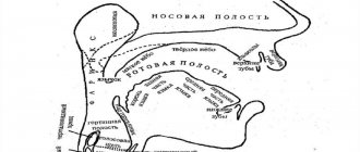
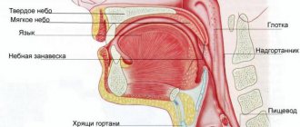
The vocal section consists of the larynx with the vocal folds located in it. The larynx is a wide, short tube consisting of cartilage and soft tissue. It is located in the front of the neck and can be felt through the skin from the front and sides, especially in thin people.
From above the larynx passes into the pharynx. From below it passes into the windpipe (trachea).
At the border of the larynx and pharynx is the epiglottis. It consists of cartilage tissue shaped like a tongue or petal. Its front surface faces the tongue, and its back surface faces the larynx. The epiglottis serves as a valve: descending during the swallowing movement, it closes the entrance to the larynx and protects its cavity from food and saliva.
Modulation of the main and additional tone of the voice
The main resonators of the human voice are the pharynx, oral cavity and nasal cavity with its paranasal sinuses, as well as the frontal cavity.
A certain timbre of voice.
The timbre is given by the cavities of the trachea and bronchi, the chest as a whole, and the laryngeal cavity. Resonators differ among individual people in shape, volume, and the characteristics of their use during speech, which gives the voice an individual timbre coloring. The soft palate and those muscles that cover the space between the nasopharynx and oropharynx take a special part in the resonance effect.
The resonators that are formed by the bones of the skull, namely the nasal cavity, the frontal cavity, do not change their volume, therefore they generate sounds in a very narrow range.
Articulatory
The volume and clarity of speech sounds are created thanks to resonators.
Resonators are located throughout the extension tube - this is everything that is located above the larynx: the pharynx, oral cavity and nasal cavity. When producing speech sounds, the extension pipe performs a dual function: a resonator and a noise vibrator.
Extension pipe.
The muscles of the tongue play a major role in the production of speech sounds. When pronouncing a single speech sound, part of the muscle fiber may be tense, while another part is relaxed. Tension of the articulatory muscle during oral speech is associated not only with the specific work of pronouncing a single sound. It bears the influence of residual stress from the utterance of the previous sound, as well as the preparatory stress associated with the utterance of the subsequent sound, which are part of the word (coarticulation). In addition, the emotional state in which the speaker is
also affects the degree of muscle tension of both the tongue and the entire speech apparatus. Thus, the muscles of the tongue experience a complex of different influences.
The tongue is a massive muscular organ. When the jaws are closed, it fills almost the entire oral cavity. The front part of the tongue is mobile, the back part is fixed and is called the root of the tongue. The movable part of the tongue is divided into the tip, the leading edge (blade), the lateral edges and the back. The complexly intertwined system of tongue muscles and the variety of their attachment points provide the ability to change the shape, position and degree of tension of the tongue within a wide range. This is very important, since the tongue is involved in the formation of all vowels and almost all consonant sounds (except labials).
Plays an important role in the formation of speech sounds. Articulation consists in the fact that the listed organs form slits, or closures, that occur when the tongue approaches or touches the palate, alveoli, teeth, as well as when the lips are compressed or pressed against the teeth.
Lower jaw, lips, teeth, hard palate, alveoli.
During quiet breathing, the soft palate is relaxed, partially closing the entrance to the oral cavity from the pharynx. During deep breathing, yawning and speech, the velum palatine rises, opening the passage into the oral cavity and, conversely, closing the passage into the nasopharynx.
Soft sky.
They take part in pronouncing all the sounds of the Russian language.
Oral cavity and pharynx.
Literature:
1. Volosovets T.V.; Overcoming general speech underdevelopment in preschool children. Educational and methodological manual / Ed. ed. - M.: V. Sekachev, 2007. - 224 p.
2. Gvozdev A. N. From first words to first grade. Diary of scientific observations. Saratov: Saratov University Publishing House, 1981
3. Speech therapy: Textbook. for students defectol. fak. ped. higher textbook institutions / Ed. Volkova L.S., Shakhovskaya S.N.;
4. Luria A. R.; Fundamentals of neuropsychology. Textbook aid for students higher textbook establishments. - M.: Publishing House, 2003. - 384 p.
5. Chirkina G.V. Programs of compensatory preschool educational institutions for children with speech impairments. – M.: Education, 2009.
The structure of the speech apparatus
The speech apparatus consists of two closely interconnected parts: the central (or regulatory) speech apparatus and the peripheral (or executive) (Fig. 1).
The central speech apparatus is located in the brain. It consists of the cerebral cortex (mainly the left hemisphere), subcortical ganglia, pathways, brainstem nuclei (primarily the medulla oblongata) and nerves going to the respiratory, vocal and articulatory muscles.
What is the function of the central speech apparatus and its departments?
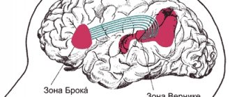
Speech, like other manifestations of higher nervous activity, develops on the basis of reflexes. Speech reflexes are associated with the activity of various parts of the brain. However, some parts of the cerebral cortex are of primary importance in the formation of speech. These are the frontal, temporal, parietal and occipital lobes of predominantly the left hemisphere of the brain (in left-handers, the right). The frontal gyrus (inferior) is a motor area and is involved in the formation of one's own oral speech (Broca's area). The temporal gyri (superior) are the speech-auditory area where sound stimuli arrive (Wernicke's center). Thanks to this, the process of perceiving someone else’s speech is carried out. The parietal lobe of the cerebral cortex is important for understanding speech. The occipital lobe is a visual area and ensures the acquisition of written speech (the perception of letter images when reading and writing). In addition, the child begins to develop speech thanks to his visual perception of the articulation of adults.
The subcortical nuclei control the rhythm, tempo and expressiveness of speech.
Conducting pathways. The cerebral cortex is connected to the speech organs (peripheral) by two types of nerve pathways: centrifugal and centripetal.
Centrifugal (motor) nerve pathways
connect the cerebral cortex with the muscles that regulate the activity of the peripheral speech apparatus. The centrifugal pathway begins in the cerebral cortex in Broca's center.
From the periphery to the center, i.e. from the region of the speech organs to the cerebral cortex, centripetal paths go.
Centripetal path
begins in proprioceptors and baroreceptors.
Proprioceptors
are found inside muscles, tendons and on the articular surfaces of moving organs.
Rice. 1. Structure of the speech apparatus: 1 - brain: 2 - nasal cavity: 3 - hard palate; 4 - oral cavity; 5 - lips; 6 - incisors; 7 - tip of the tongue; 8 - back of the tongue; 9 - root of tongue; 10 - epiglottis: 11 - pharynx; 12 —
larynx; 13 - trachea; 14 - right bronchus; 15 - right lung: 16 - diaphragm; 17 - esophagus; 18 - spine; 19 - spinal cord; 20 - soft palate
CHAPTER 1 Basic mechanisms of oral speech
1.1 Anatomical and physiological mechanisms of speech
Speech is a product of human mental activity and the result of a complex interaction of different brain structures. The implementation of oral speech occurs thanks to the coordinated work of the peripheral motor apparatus, which is provided by the central nervous system.
The respiratory, phonatory and articulatory sections of the peripheral speech apparatus are involved in speech production.
The respiratory section of the peripheral speech apparatus forms the energetic basis of speech, providing the so-called speech breathing. Anatomically, this section is represented by the chest, lungs, intercostal muscles and muscles of the diaphragm. The lungs provide a certain subglottic air pressure. It is necessary for the functioning of the vocal folds, voice modulations and changes in its tonality.
During physiological breathing (i.e., outside of speech), inhalation occurs actively due to contraction of the respiratory muscles, and exhalation occurs relatively passively due to the lowering of the chest walls and the elasticity of the lungs. The inhalation and exhalation phases at rest differ little in duration. According to the method of preferential expansion of the thoracic cavity, physiological breathing is divided into various types: 1) costal (chest); 2) abdominal; 3) mixed (thoracic-abdominal). In turn, costal breathing comes in three varieties: a) clavicular; b) upper costal; c) lower costal. Clavicular and upper costal breathing refers to irrational breathing methods, since the expansion of the chest is limited due to the low mobility of the costal walls. With abdominal breathing, the tidal volume does not differ significantly from that with lower costal breathing, but the respiratory movements are more flexible. More rational is thoraco-abdominal breathing, which is often called diaphragmatic in practice. This type of breathing ensures not only a sufficient volume of air, but also optimal plasticity of respiratory movements. This type of breathing is most adequate for phonation.
During speech, the functional significance of the exhalation phase increases significantly. Before speaking, a quick and deeper breath is usually taken than at rest. Speech inhalation is carried out through the nose and mouth, and in the process of speech exhalation, the air flow goes only through the mouth. “Speech” inspiration is characterized by the presence of a certain volume of air that can ensure maintenance of ligamentous pressure. A rational way of using the air stream is of great importance for voicing a statement. The exhalation time is extended as much as the sound of the voice is necessary during the continuous pronunciation of an intonation-logically completed segment of the utterance (i.e., syntagm).
The phonatory section of the peripheral speech apparatus is anatomically represented by the larynx and its vocal folds. Outside of speech, the folds are moved apart. During phonation, the vocal folds tense, close and produce oscillatory movements. It is the oscillatory movements of the vocal folds that generate sound waves.
The frequency and power characteristics of the human voice are a reflection of the amplitude and frequency of vibration of the vocal folds.
The main and additional tones of the voice are modulated by a system of resonators. The main resonators of the human voice are the pharynx, oral cavity and nasal cavity with its paranasal sinuses, as well as the frontal cavity. In addition, the cavities of the trachea and bronchi, the chest as a whole, and the laryngeal cavity give a certain timbre to the voice. Resonators differ among individual people in shape, volume, and the characteristics of their use during speech, which gives the voice an individual timbre coloring.
The soft palate and those muscles that cover the space between the nasopharynx and oropharynx take a special part in the resonance effect.
The resonators that are formed by the bones of the skull, namely the nasal cavity, the frontal cavity, do not change their volume, therefore they generate sounds in a very narrow range.
The frequency range of the human voice is measured in hertz. The frequency range of the spoken voice is only 1/10 of the total voice range. In men, the frequency range of the voice is 80-150 Hz, in women - 120-400 Hz, in children it is much higher. Since human hearing is unequally sensitive to sounds of different frequencies, the perceived loudness of a voice depends not only on the absolute strength, but also on its frequency characteristics. High-pitched voices feel louder.
The color of the voice reflects the emotional state of the speaker and even the mental state of the individual in the broadest sense of the word.
There are timbre differences in the vocal range, which, by analogy with musical instruments, are called vocal registers. There are three registers in the human voice: chest, head and middle (mixed).